Effect of Steaming and Ultrasound Treatment on Sulforaphane and Allyl Isothiocyanate Content of Brussels Sprouts (Brassica oleracea var. gemmifera)
Abstract
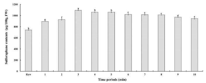
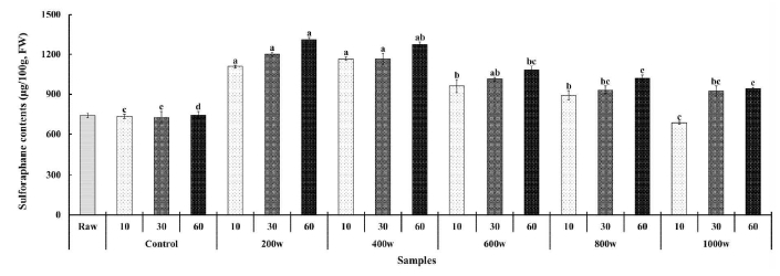
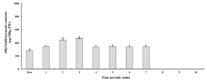
The objective of this study was to investigate the effect of different durations of steaming (ranging from 1 to 10 min) and ultrasound treatments at various intensities (200 w, 400 w, 600 w, 800 w, or 1,000 w) and durations (10, 30, or 60 min) on the levels of sulforaphane (SFN) and allyl isothiocyanates (AITC) in Brussels sprouts. The SFN content continued to increase up to 3 min of steaming time, after which it decreased. However, even after this decrease, the content remained significantly higher (p<0.05) than that of the raw sample, reaching its maximum value of 1,095.57±7.24 μg/100 g fresh weight (FW) at 3 min of steaming time. In response to the increase in ultrasonic power intensity, the SFN content exhibited an upward trend, followed by a decrease. The highest SFN content was observed at an ultrasonic power intensity of 200 w and a processing time of 60 min (1,312.78±14.02 μg/100 g FW). Additionally, the AITC content continued to increase up to 3 min of steaming time (466.31±3.34 μg/100 g FW), followed by a subsequent decrease, maintaining a consistent level from 4 to 7 min of steaming time. With the ultrasonic treatment, the AITC content increased until an intensity of 400 w was reached, after which it declined beyond 600 w. The treatment conditions yielding the highest content were identified as 200 w and 60 min. Thus, this study demonstrates that both steaming and ultrasonic treatment can affect myrosinase enzyme inactivation and suggests that ultrasonic treatment may yield higher increases in SFN and AITC content for Brussels sprouts compared to steaming.
Keywords:
brussels sprouts, steaming, ultrasound, sulforaphane, allyl isothiocyanatesINTRODUCTION
Brussels sprouts (Brassica oleracea var. gemmifera) are similar in appearance to ordinary cabbage (Brassica oleracea var. capitata), but they are also known as “Brassican cabbage” or “mini cabbage” because each sprout is attached to a long stem, resembling a bell (Hwang ES 2019). Brussels sprouts are widely cultivated in Europe and the United States, and are a cruciferous vegetable that is popularly consumed in winter due to their excellent taste, quality, and storage properties (Mun W et al 2014). Although brussels sprouts were first introduced to Korea in the 1990s and cultivated in small quantities in Jeju, with the recent surge in their popularity, the cultivated area has significantly increased. Brussels sprouts are a type of cruciferous vegetable that are known to be rich in sulfur-containing compounds called glucosinolates (Fenwick GR et al 1983). When these compounds are hydrolyzed in the presence of water by the enzyme myrosinase (β-thioglucosidase glucohydrolase, EC 3.2.1.147), they form isothiocyanates (ITCs) (Hecht SS 2000). There are many types of glucosinolates (GSL) found in cruciferous vegetables, including brussels sprouts, and each type produces different ITCs such as sulforaphane (SFN), phenylethyl isothiocyanates (PEITC), and allyl isothiocyanates (AITC), as well as indole compounds (Yang G et al 2010). ITCs are considered to be potentially bioactive and are currently being studied for their anticarcinogenic properties, including their ability to induce detoxification enzymes and inhibit genes that promote tumor formation (Tanii H et al 2008).
Ultrasound processing is a new and environmentally friendly technology being adopted by the food industry for different purposes, including tissue disruption and enzyme inactivation (Fu X et al 2020). Ultrasound uses acoustic waves with frequencies beyond the human audible range, and its effects on food are due to the cavitation phenomenon, which is the formation and collision of bubbles accompanied by high temperature and pressure, causing disruption to cell walls and membranes, and reducing enzyme activity by breaking hydrogen interactions and van der Waals forces (Iqbal A et al 2019). The use of ultrasound in biotechnological processes has garnered attention from various research groups (Hwang SH & Koo YM 2001; Lee KJ & Row KH 2006). The ultrasonication method is widely used on a laboratory scale due to its simplicity and the lack of need for complex equipment or extensive technical training. Ultrasound irradiation can alter the structure and function of biological molecules through several mechanisms, including heat, chemical effects, and acoustically induced cavitational activity. Additionally, mechanical effects caused by shear stress developed from shock waves can lead to the inactivation of biomolecules during ultrasonication (Suslick KS 1990). Ultrasound waves have been found to have the potential to affect enzymes. The effects of ultrasonic power on enzymes can be categorized into three basic types: (1) Aiding in biological reactions, (2) causing a decrease in enzyme activity for many enzymes in vitro, and (3) in some cases, increasing the activity of free enzymes instead of deactivating them. Several studies have shown that ultrasound can enhance enzyme-catalyzed reactions. For example, ultrasound has been found to improve the rate of enzyme-catalyzed hydrolysis of starch and sucrose via alpha-amylase (Barton S et al 1996; Apar DK et al 2006), and the rate of hydrolysis of milk lactose under sonication (Sener N et al 2006). While there are many reports of ultrasound decreasing enzyme activity (López P et al 1994; López P & Burgos J 1995; López P et al 1998), only a limited number of studies have found that ultrasound can increase activity for free enzymes in vitro. Remarkably, at low acoustic power, some enzymes, whether supported on porous silica gel or free, are not deactivated, such as alpha-amylase (EC 3.2.1.1) and glucoamylase (EC 3.2.1.3) (Schmidt P et al 1987).
Different studies have reported that mild ultrasound irradiation can increase the activity of free enzymes, and that in some cases, the rate of activity increases at lower intensities followed by a decrease at higher intensities, particularly for alpha-chymotrypsin on casein (Schmidt P et al 1987). Conversely, high intensities of ultrasound have been found to decrease the activity of many enzymes in vitro, likely due to changes in the structure and function of biological molecules. However, in some cases, low-intensity ultrasound can actually increase the activity of free enzymes instead of deactivating them. The intensity level of ultrasound is therefore a key factor in determining the activity or inactivity of many enzymes. Additionally, it appears that enzymes, which are essential to biochemical reactions, increase their activity with an appropriate amount of ultrasound. Generally, combining ultrasonication with other treatments is more effective at enhancing inactivation efficacy. Some authors have explored the efficacy of combining heat with power ultrasound (thermosonication), and they found that enzyme inactivation was greater with thermosonication than the sum of the effects of heat and ultrasound acting independently, making it a more efficient option in terms of treatment time and energy consumption compared to using either treatment individually (Ordóñez JA et al 1984). Currently, there have been no studies conducted to examine the impact of steaming and ultrasound processing on the levels of SFN and AITC in brussels sprouts. Therefore, the objective of this study is to investigate the effects of steaming and ultrasound processing on SFN and AITC content of brussels sprouts.
MATERIALS AND METHODS
1. Chemicals and Reagents
Sulforaphane, allyl isothiocyanate, HPLC-grade methanol and acetonitrile were purchased from Sigma-Aldrich Co. (St. Louis, MO, USA). Bakerbond SPE silica gel (SiOH) 3 mL disposable columns were from J.T. Baker (Phillipsburg, NJ, USA). Dichoromethane, ethylacetate, methanol, methanol, and ammonium acetate were obtained from Merck (Darmstadt, Germany), unless mentioned otherwise.
2. Sample Preparation
Brussels sprouts were purchased from local market. They were washed with water and dried with paper towels. The impact on the levels of SFN and AITC in brussels sprouts after steaming and ultrasonication relative to untreated brussels sprouts were studied. For steaming, after adding 1 L of distilled water to the electric steamer (HD-9132, Philips Co, Seoul, Korea) and heated it until the internal temperature reached 100℃. Then, 1 kg of brussels sprouts was placed into the steamer for the heat treatment. The heat treatment was performed for various durations, ranging from 1 to 10 min. At one-minute intervals, 100 g portions of the brussels sprouts were removed and the content SFN and AITC were determined immediately after steaming treatment. For ultrasound treatment, after washing 1 kg of brussels sprouts with distilled water, it immersed them in an ultrasound equipment (powersonic 440, Hwashin Co, Seoul, Korea) containing 35 L of distilled water. The ultrasound equipment operated at a frequency of 40 kHz, while the temperature of the chamber was set to 40℃. The brussels sprouts underwent ultrasound treatment at different intensities (200 w, 400 w, 600 w, 800 w, or 1,000 w) and for varying durations (10, 30, or 60 min). To serve as a control group, another 1 kg of brussels sprouts was placed in a thermostatic water bath at 40℃ and treated for the same durations (10, 30, or 60 min). The content SFN and AITC were determined immediately after ultrasound treatment.
3. Extraction and Purification of SFN from Brussels Sprouts
Samples were weighed out in a beaker of 50 mL (1 g fresh sample), then 4 mL of acidic water (pH 6) was added, and the mixture was incubated at 45±2℃ in water bath for 2.5 h. At this step, the evaporation of the water was observed to have decreased the extract by about 70% from the initial weight of the mixture (sample+acidic water). Sulforaphane was extracted with 20 mL of dichloromethane by vortexing for 1 min and stored at room temperature during 1 h. Then, the resulting solution was filtered through Whatman no. 41 paper. The sulforaphane was purified with the method designed by Bertelli D et al (1998), using Bakerbond SPE silica gel (SiOH) 3 mL disposable columns. Prior to use, the silica gel cartridge was conditioned with 3 mL of dichoromethane. Sulforaphane was extracted by passing 20 mL of organic extract through the cartridge, washing the cartridge with 3 mL of ethylacetate (which was then discarded) and eluting the sulforaphane with 3 mL of methanol. The methanol extract was evaporated to dryness in a vacuum oven at 45℃ for 2 h, and redissolved with 2 mL of acetonitrile. The resulting solution was vortexed for 30 s and filtered with a membrane of 0.45 μm. A 20 μL sample of this solution was injected onto the column of the HPLC system (Agilent 1260 Infinity Quaternary liquid chromatograph, Hewlett Packard, Wilmington, NC, USA). All samples were analyzed five times.
4. SFN Analysis
Analyses of SFN were performed using an Agilent 1260 infinity quaternary liquid chromatograph (Hewlett Packard, Wilmington, NC, USA) equipped with a quaternary gradient pump and multiple wavelength detector operating at 202 nm. The samples were separated on a Zorbax Eclipse XDB-C18 column (4.6 mm × 150 mm, 5 μm; Agilent Technologies, Inc., Santa Clara, CA, USA) at 36℃ with a sample injection volume of 10 μL. The analysis were carried out isocratically at a flow rate of 0.6 mL/min, employing as the mobile phase a mixture of acetonitrile : ultrapure water (30 : 70, v/v). Data analysis was performed using Chemstation software (Hewlett Packard, Wilmington, NC, USA).
5. Extraction of AITC from Brussels Sprouts
The mixtures were homogenized on an orbital shaker (VWR Mini Shaker, VWR, Darmstadt, Germany) at 450 rpm for 10 min and then sonicated for 30 min. The mixtures were subsequently centrifuged at 20,000 rpm at 7℃ for 10 min. A 2 mL syringe was used to remove the supernatant and the extract was then filtered using a 0.45 µm RC membrane filter (Phenomenex, Aschaffenburg, Germany). Five replicates of each sample were analyzed.
6. AITC Analysis
Analyses of AITC were performed using an Agilent 1260 infinity quaternary liquid chromatograph (Hewlett Packard, Wilmington, NC, USA) with a multiple wavelength detector (MWD) operating at 242 nm. Chromatographic separations were performed on a Supelcosil LC-18 RP column (4.6 × 150 mm, 5 µm, Sigma-Aldrich, St. Louis, MO, USA) using methanol and 0.1 N ammonium acetate (NH4OAc) (3:97; v/v) as the mobile phase. The column temperature was 30℃, and the flow rate was 1 mL/ min. Data analysis was performed using Chemstation software (Hewlett Packard, Wilmington, NC, USA).
7. Statistical Analysis
The experimental data were subjected to analysis of variance (ANOVA). Significant differences between the mean values, as determined from measurements carried out in five replicate tests (i.e., p<0.05), were obtained by Duncan’s multiple-range test using statistical analysis software (SPSS 20.0, IBM Inc., NY, USA).
RESULTS AND DISCUSSION
1. Linearity
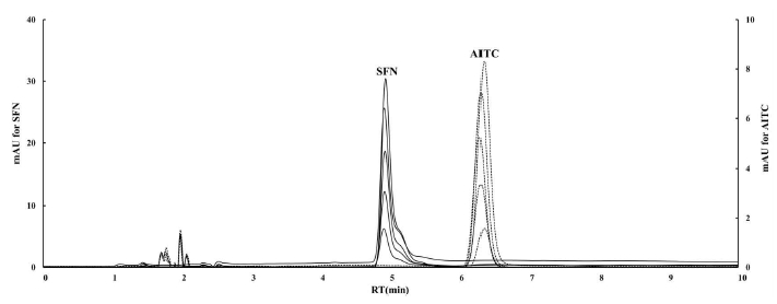
Linear regression analysis was conducted to assess the linearity of the obtained data. The response of SFN and AITC exhibited linearity within the concentration range of 2—10 mg/L. The linear regression equations for SFN and AITC were determined as y=7.5392x—0.2858 with an r² value of 0.9991 and y=2.012x—0.06 with an r² value of 0.9964, respectively. The calibration plot is illustrated in Fig. 1, and the chromatogram is presented in Fig. 2. The coefficient of determination's acceptance value should not fall below 0.99, and the determined values, ranging from 0.993 to 0.9998, satisfied this criterion. The detection limits for SFN and AITC were both established at 0.05 mg/L.
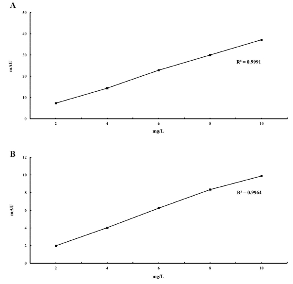
Calibration curve measured at 202 nm with r2=0.0991 for SFN (A) and 242 nm with r2=0.0964 for AITC (B).
2. Effect of Steaming Treatment on SFN Content of Brussels Sprouts
The effect of steaming treatment on SFN content from brussels sprouts was analyzed was studied for different processing times, ranging from 1 to 10 min (Fig. 3). The initial content of SFN in unprocessed samples was found to be 741.09±5.89 µg/100 g FW. The content of SFN increased up to 3 min of steaming time, but decreased thereafter, reaching a level of 946.85±10.76 µg/100 g FW at 10 min. However, this content still showed a significantly (p<0.05) higher amount compared to the raw sample. Moreover, the SFN content exhibited the lowest value of 894.65±1.32 µg/100 g FW at 1 min, and the highest value of 1,095.57±7.24 µg/100 g FW at 3 min of steaming time. The rise in sulforaphane content can be attributed to the application of high temperatures, which likely led to a considerable reduction in myrosinase activity. However, the remaining glucoraphanin can still be converted into sulforaphane due to the enzymatic activity of the microbiota (Fahey JW et al 2012). Remarkably, sulforaphane demonstrated greater stability than myrosinase (Wu Y et al 2018), allowing it to accumulate despite ongoing myrosinase hydrolysis and limited thermal degradation. As the cooking time increased, the content of glucoraphanin experienced a notable decrease, and elevated temperatures accelerated the generation of specific volatile compounds from sulforaphane (Jin Y et al 1999). This series of events led to the decline in sulforaphane content. Notably, the sulforaphane content reached its peak at the 3 min of the steaming process (Fig. 3). Therefore, this finding can play a crucial role in determining the optimal cooking time to retain a high level of sulforaphane.
3. Effect of Ultrasound Treatment on SFN Content of Brussels Sprouts
The effect of ultrasound treatment on the formation of sulforaphane in brussels sprouts was examined at different intensities of ultrasound treatment ranging from 200 to 1,000 w and three different processing durations (10, 30, or 60 min) (Fig. 4). The content of sulforaphane exhibited an increasing trend followed by a decrease in response to the increase of ultrasonic power intensity. The high content of SFN was observed at ultrasonic power intensities of 200 w (1,108.43—1,312.78 µg/100 g FW) and 400 w (1,166.00—1,274.48 µg/100 g FW), while it decreased for power intensities exceeding 600 w. Notably, even in comparison to both the raw and control groups, the content of sulforaphane remained notably higher. Additionally, in all treatment groups of different power intensities, the content of sulforaphane increased as processing time extended. The treatment group with the highest sulforaphane content was observed at an ultrasonic power intensity of 200 w and a processing time of 60 min (1,312.78±14.02 µg/100 g FW). However, no significant difference was found between this group and the sample treated with an ultrasonic power intensity of 400 w and a processing time of 60 min (1,274.48±17.08 µg/100 g FW). The application of ultrasound processing in brussels sprouts has been relatively underexplored until now. Briones-Labarca V et al (2015) found that ultrasound-assisted extraction led to a 5-fold increase in SFN recovery from Chilean papaya seeds in a methanol solution. Pongmalai P et al (2017) noted that ultrasound-assisted extraction significantly improved glucoraphanin extractability (by 1.8-fold) from steamed cabbage leaves compared to fresh cabbage when using methanol as a solvent. Addition, ultrasound treatment as a mild heating treatment might induce more cell lysis, resulting in the diffusion of greater amounts of glucosinolates and myrosinase, thus promoted the formation of sulforaphane (Nugrahedi PY et al 2015). In the present study, ultrasound treatment significantly improved sulforaphane content in brussels sprouts compared with the control (Fig. 4). Besides, Olivero T et al (2014) reported that ultrasound processing lowers the activation energy required for myrosinase inactivation. This could represent significant energy savings in an industrial process, constituting a more efficient blanching process in comparison with traditional blanching. Myrosinase inactivation may be useful to preserve glucoraphanin in the vegetable tissue if the objective is extracting the precursor of SFN, glucoraphanin, to conduct the hydrolysis exogenously under controlled conditions. This will prevent the formation of undesirable compounds such as nitriles and isothionitriles which compete with SFN formation, thus maximizing the conversion of glucoraphanin in SFN. Therefore, ultrasonic treatment not only affects the inactivation of myrosinase, leading to increased sulforaphane content in brussels sprouts, but also offers the potential to achieve higher levels than by thermal processing, thus reducing the temperature necessaries to achieve the desired denaturation.
4. Effect of Steaming Treatment on AITC Content of Brussels Sprouts
The effect of steaming treatment on AITC content from brussels sprouts was analyzed was studied for different processing times, ranging from 1 to 10 min (Fig. 5). The initial AITC content in the raw sample was found to be 279.80±2.13 µg/100 g FW. As the steaming progressed, The AITC content increased up to 3 min of steaming time (466.31±3.34 µg/100 g FW). Subsequently, there was a decrease in AITC content, while the level remained consistent from 4 to 7 min of steaming time. However, AITC was not detectable after 8 min of steaming time. AITC is a compound found in plants, stored in a stable form as its precursor called sinigrin (allyl glucosinolate or 2-propenyl glucosinolate), which belongs to the aliphatic glucosinolate category. It’s notably abundant in vegetables like brussels sprouts, broccoli, and mustard seeds derived from Brassica nigra. These plants owe their characteristic pungency to AITC (Ishida M et al 2014). Myrosinase, an enzyme, acts on sinigrin by breaking down its glucose component, forming an intermediate aglycone. This intermediate structure is inherently unstable and rearranges itself spontaneously to yield AITC (Krul C et al 2002). The isothiocyanates formed through glucosinolate hydrolysis are unstable and can easily transform into thiocyanate ions and indole3-carbinol. Moreover, steaming has the potential to preserve some level of myrosinase activity, with myrosinase remaining active for up to 2 min of heating, but showing a significant 90% decline after 7 min (Fan Y et al 2016). Opting for shorter steaming durations could result in a greater formation of ITCs compared to nitriles. This effect is due to the inactivation of the epithiospecifier protein, which occurs at a lower temperature than the inactivation of myrosinase, as reported in the case of broccoli (Green DR & Llambi F 2015). Therefore, the complete impact of vegetables containing glucosinolates can be accurately evaluated by specifically examining how different cooking conditions affect both the glucosinolate content and the activity of myrosinase.
5. Effect of Ultrasound Treatment on AITC Content of Brussels Sprouts
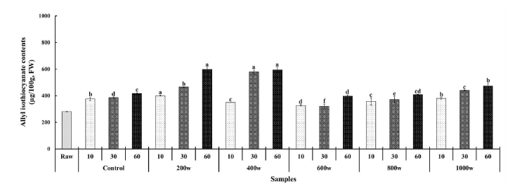
The effect of ultrasound treatment on the formation of AITC in brussels sprouts was examined at different intensities of ultrasound treatment ranging from 200 to 1,000 w and three different processing durations (10, 30, or 60 min) (Fig. 6). The initial AITC content measured 279.80±2.13 µg/100 g FW in the raw sample, while the control group exhibited levels ranging from 375.63±12.13 µg/100 g FW to 416.32±5.23 µg/100 g FW, dependent on processing time. Within the ultrasonic treatment groups, the AITC content varied with ultrasonic power intensity. It demonstrated an ascending pattern until reaching an ultrasonic intensity of 400 w, after which it declined beyond 600 w. The treatment condition yielding the highest content was identified in the 200 w, 60 min treatment group, showing a content of 595.74±14.79 µg/100 g FW. Furthermore, the AITC content increased in all treatment groups with increasing ultrasonic treatment time. As ultrasound treatment induces cell membrane disruption (Nowacka M & Wedzik M 2016), it was expected that higher levels of AITC would result from ultrasound application due to the decompartmentalization of enzymes (myrosinase) and substrates (glucosinolates) from cell organelles, allowing their interaction. However, in this study, no significant difference was observed in the AITC content according to the ultrasonic power intensity. These findings suggest that myrosinase released from ultrasound-treated brussels sprouts was partially inactivated by the applied ultrasound conditions. It is established that ultrasound can deactivate enzymes by modifying the protein’s chemical structure, with its efficacy contingent upon various factors such as ultrasound power, temperature, time, frequency, and pH (Islam MN et al 2014). Therefore, it is possible that the ultrasound treatment applied in this study partially inactivated myrosinase, thereby impeding the hydrolysis of glucosinolates.
CONCLUSION
The objective of this study was to explore the effect of different durations of steaming (ranging from 1 to 10 min) and ultrasound treatments at various intensities (200 w, 400 w, 600 w, 800 w, or 1,000 w) and durations (10, 30, or 60 min) on the levels of SFN and AITC in brussels sprouts. The SFN and AITC content exhibited the highest value at 3 min of steaming time. And the highest SFN and AITC content was observed at an ultrasonic power intensity of 200 w and a processing time of 60 min. Therefore, in this study, it has been demonstrated that both blanching and ultrasonic treatment can influence the enzyme inactivation of myrosinase. Furthermore, for brussels sprouts, it has been suggested that ultrasonic treatment could achieve higher levels of SFN and AITC content increase compared to steaming treatment. However, in future research, it is estimated necessary to explore the optimal conditions involving various factors such as heat treatment, ultrasonic treatment, processing temperature, and processing time to achieve high levels of SFN and AITC content.
References
-
Apar DK, Turhan M, Özbek B (2006) Enzymatic hydrolysis of starch by using a sonifier. Chem Eng Commun 193(9): 1117-1126.
[https://doi.org/10.1080/00986440500354424]

-
Barton S, Bullock C, Weir D (1996) The effects of ultrasound on the activities of some glycosidase enzymes of industrial importance. Enzyme Microb Technol 18(3): 190-194.
[https://doi.org/10.1016/0141-0229(95)00092-5]

-
Bertelli D, Plessi M, Braghiroli D, Monzani A (1998) Separation by solid phase extraction and quantification by reverse phase HPLC of sulforaphane in broccoli. Food Chem 63(3): 417-421.
[https://doi.org/10.1016/S0308-8146(98)00052-1]

-
Briones-Labarca V, Plaza-Morales M, Giovagnoli-Vicuña C, Jamett F (2015) High hydrostatic pressure and ultrasound extractions of antioxidant compounds, sulforaphane and fatty acids from Chilean papaya (Vasconcellea pubescens) seeds: Effects of extraction conditions and methods. LWT-Food Sci Technol 60(1): 525-534.
[https://doi.org/10.1016/j.lwt.2014.07.057]

-
Fahey JW, Wehage SL, Holtzclaw WD, Kensler TW, Egner PA, Shapiro TA, Talalay P (2012) Protection of humans by plant glucosinolates: Efficiency of conversion of glucosinolates to isothiocyanates by the gastrointestinal microflora. Cancer Prev Res 5(4): 603-611.
[https://doi.org/10.1158/1940-6207.CAPR-11-0538]

-
Fan Y, Yang F, Cao X, Chen C, Zhang X, Zhang X, Lin W, Wang X, Liang C (2016) Gab1 regulates SDF-1-induced progression via inhibition of apoptosis pathway induced by PI3K/AKT/Bcl-2/BAX pathway in human chondrosarcoma. Tumor Biol 37(1): 1141-1149.
[https://doi.org/10.1007/s13277-015-3815-2]

-
Fenwick GR, Heaney RK, Mullin WJ (1983) Glucosinolates and their breakdown products in food and food plants. Crit Rev Food Sci Nutr 18(2): 123-194.
[https://doi.org/10.1080/10408398209527361]

-
Fu X, Belwal T, Cravotto G, Luo Z (2020) Sono-physical and sono-chemical effects of ultrasound: Primary applications in extraction and freezing operations and influence on food components. Ultrason Sonochem 60: 104726.
[https://doi.org/10.1016/j.ultsonch.2019.104726]

-
Green DR, Llambi F (2015) Cell death signaling. Cold Spring Harb Perspect Biol 7(12): a006080.
[https://doi.org/10.1101/cshperspect.a006080]

-
Hecht SS (2000) Inhibition of carcinogenesis by isothiocyanates. Drug Metab Rev 32(3-4): 395-411.
[https://doi.org/10.1081/DMR-100102342]

-
Hwang ES (2019) Effect of cooking methods on bioactive compound contents and antioxidant activities of Brussels sprouts. J Korean Soc Food Sci Nutr 48(10): 1061-1069.
[https://doi.org/10.3746/jkfn.2019.48.10.1061]

- Hwang SH, Koo YM (2001) Effects of operation and design parameters on the recovery of microorganisms and particles in ultrasonic sedimentation. Korean Chem Eng Res 39(6): 788-793.
-
Iqbal A, Murtaza A, Hu W, Ahmad I, Ahmed A, Xu X (2019) Activation and inactivation mechanisms of polyphenol oxidase during thermal and non-thermal methods of food processing. Food Bioprod Process 117: 170-182.
[https://doi.org/10.1016/j.fbp.2019.07.006]

-
Ishida M, Hara M, Fukino N, Kakizaki T, Morimitsu Y (2014) Glucosinolate metabolism, functionality and breeding for the improvement of Brassicaceae vegetables. Breed Sci 64(1): 48-59.
[https://doi.org/10.1270/jsbbs.64.48]

-
Islam MN, Zhang M, Adhikari B (2014). The inactivation of enzymes by ultrasound-A review of potential mechanisms. Food Rev Int 30(1): 1-21.
[https://doi.org/10.1080/87559129.2013.853772]

-
Jin Y, Wang M, Rosen RT, Ho CT (1999) Thermal degradation of sulforaphane in aqueous solution. J Agric Food Chem 47(8): 3121-3123.
[https://doi.org/10.1021/jf990082e]

-
Krul C, Humblot C, Philippe C, Vermeulen M, Van Nuenen M, Havenaar R, Rabot S (2002) Metabolism of sinigrin (2-propenyl glucosinolate) by the human colonic microflora in a dynamic in vitro large-intestinal model. Carcinogenesis 23(6): 1009-1016.
[https://doi.org/10.1093/carcin/23.6.1009]

-
Lee KJ, Row KH (2006) Enhanced extraction of isoflavones from Korean soybean by ultrasonic wave. Korean J Chem Eng 23(5): 779-783.
[https://doi.org/10.1007/BF02705927]

-
López P, Burgos J (1995) Lipoxygenase inactivation by manothermosonication: Effects of sonication on physical parameters, pH, KCl, sugars, glycerol, and enzyme concentration. J Agric Food Chem 43(3): 620-625.
[https://doi.org/10.1021/jf00051a012]

-
López P, Sala FJ, Fuente JLDL, Condon S, Raso J, Burgos J (1994) Inactivation of peroxidase, lipoxygenase, and polyphenol oxidase by manothermosonication. J Agric Food Chem 42(2): 252-256.
[https://doi.org/10.1021/jf00038a005]

-
López P, Vercet A, Sánchez AC, Burgos J (1998) Inactivation of tomato pectic enzymes by manothermosonication. Zeitschrift für Lebensmitteluntersuchung und -forschung A 207(3): 249-252.
[https://doi.org/10.1007/s002170050327]

- Mun W, Kim JG, Lee JW (2014) Cabbage and Vegetable. Korea National Open University Publishing, Korea. p 355.
-
Nowacka M, Wedzik M (2016) Effect of ultrasound treatment on microstructure, colour and carotenoid content in fresh and dried carrot tissue. Appl Acoust 103: 163-171.
[https://doi.org/10.1016/j.apacoust.2015.06.011]

-
Nugrahedi PY, Verkerk R, Widianarko B, Dekker M (2015) A mechanistic perspective on process-induced changes in glucosinolate content in brassica vegetables: A review. Crit Rev Food Sci 55(6): 823-838.
[https://doi.org/10.1080/10408398.2012.688076]

-
Oliviero T, Verkerk R, Van Boekel MAJS, Dekker M (2014) Effect of water content and temperature on inactivation kinetics of myrosinase in broccoli (Brassica oleracea var. italica). Food Chem 163(15): 197-201.
[https://doi.org/10.1016/j.foodchem.2014.04.099]

-
Ordoñez JA, Sanz B, Hernandez PE, Lopez-Lorenzo P (1984) A note on the effect of combined ultrasonic and heat treatments on the survival of thermoduric streptococci. J Appl Bacteriol 56(1): 175-177.
[https://doi.org/10.1111/j.1365-2672.1984.tb04711.x]

-
Pelosi C, Chiron F, Dubs F, Hedde M, Ponge JF, Salmon S, Cluzeau D, Nélieu S (2014) A new method to measure allyl isothiocyanate (AITC) concentrations in mustard-Comparison of AITC and commercial mustard solutions as earthworm extractants. Appl Soil Ecol 80: 1-5.
[https://doi.org/10.1016/j.apsoil.2014.03.005]

-
Pongmalai P, Devahastin S, Chiewchan N, Soponronnarit S (2017) Enhancing the recovery of cabbage glucoraphanin through the monitoring of sulforaphane content and myrosinase activity during extraction by different methods. Sep Purif Technol 174(1): 338-344.
[https://doi.org/10.1016/j.seppur.2016.11.003]

-
Schmidt P, Rosenfeld E, Millner R, Czerner R, Schellenberger A (1987) Theoretical and experimental studies on the influence of ultrasound on immobilized enzymes. Biotechnol Bioeng 30(8): 928-935.
[https://doi.org/10.1002/bit.260300803]

-
Sener N, Apar DK, Özbek B (2006) A modelling study on milk lactose hydrolysis and beta-galactosidase stability under sonication. Process Biochem 41(7): 1493-1500.
[https://doi.org/10.1016/j.procbio.2006.02.008]

-
Suslick KS (1990) Sonochemistry. Science 247(4949): 1439-1445.
[https://doi.org/10.1126/science.247.4949.1439]

- Tanii H, Higashi T, Nishimura F, Higuchi Y, Saijoh K (2008) Effects of cruciferous allyl nitrile on phase 2 antioxidant and detoxification enzymes. Med Sci Monit 14(10): BR189-BR192.
-
Wu Y, Shen Y, Wu X, Zhu Y, Mupunga J, Bao W, Huang J, Mao J, Liu S, You Y (2018) Hydrolysis before stir-frying increases the isothiocyanate content of broccoli. J Agric Food Chem 66(6): 1509-1515.
[https://doi.org/10.1021/acs.jafc.7b05913]

-
Yang G, Gao YT, Shu XO, Cai Q, Li GL, Li HL, Ji BT, Rothman N, Dyba M, Xiang YB, Chung FL, Chow WH, Zheng W (2010) Isothiocyanate exposure, glutathione S-transferase polymorphisms, and colorectal cancer risk. Am J Clin Nutr 91(3): 704-711.
[https://doi.org/10.3945/ajcn.2009.28683]

